Overview
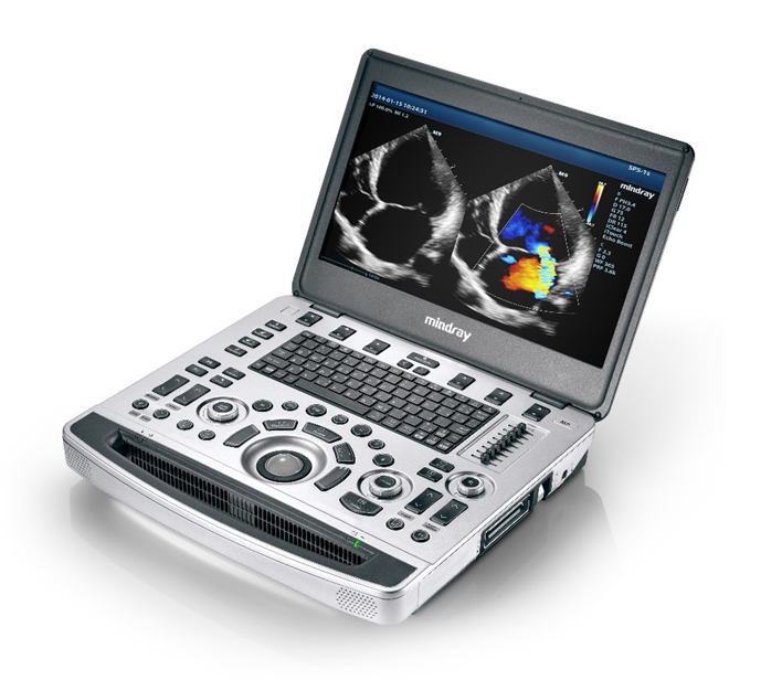
Based on Mindray's new generation ultrasound platform, mQuadro, M9Vet has raised the industry standards to an all new level. Advanced signal transmission and reception processors provide highly sensitive and accurate echo detection. Innovative transducer technologies allow for better penetration, higher resolution, greatly enhancing your diagnostic experience.
Performance
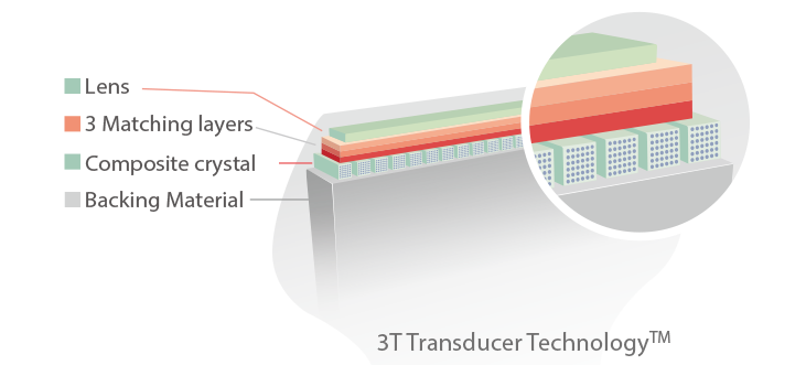
3T Transducer Technology with Single Crystal
Providing sharper images, all probes compatible with the M9Vet come equipped with Mindray’s unique 3T transducer technology. Enhanced with single-crystal technology, M9Vet offers better penetration and dynamic color flow, especially during difficult patients scanning.
Echo Boost™
Mindray’s unique adaptive signal processing technology with intelligent echo detection is designed to utilize the native signal-to-noise information, to enhance the weak echo signals. At the same time, they are suppressing the surrounding clutter noise, providing more balanced image brightness and improved visualization of myocardium tissue layers.
Tissue Tracking with Quantitative Analysis
The TT QA functionality on M9Vet allows for a simple, quick, and non-invasive solution to evaluation left ventricular wall motion abnormalities. Supported by Mindray’s patented 3T technology with single crystal, M9Vet significantly improves the tracking accuracy and effectiveness, controlling the image drift caused by the probe movement and respiration. With the added unique benefit of on-site analyses, the TT QA on M9Vet can be performed at the bedside, saving time and making complicated diagnoses much simpler.
Natural Touch Elastography
The latest Mindray patented technology- natural touch elastography, reduces user operation technique dependency. It also improves operator’s reproducibility for higher clinical utility.
- Higher stiffness sensitivity
- Good stability and reproducibility
UWN+ Contrast Imaging (Ultra-Wideband Non-linear)
M9Vet’s most significant advantage lies in its ability to support Mindray’s patented technology to enhance contrast imaging capability. UWN+ contrast imaging enables M9Vet to detect and utilize both 2nd harmonic and non-linear fundamental signals, generating images with significant enhancements.
- More sensitive to weak signal leading to reduced agent dosage
- Longer agent duration with lower MI requirement
PSHI™(Phase Shift Harmonic Imaging)
The purified Harmonic Imaging for better contrast resolution provides clearer images with excellent resolution and less noise.
Tissue Harmonic Imaging (THI)
THI significantly enhances contrast resolution and improves image quality by utilizing second harmonics generated from tissue boundary layers, especially for technically challenging subjects.
iBeam™
Gain improved image quality based on auto structure detection.
- Sharper & Continuous Edges
- Smooth Uniform Tissues
- Cleaner ‘no echo areas’
Multi-Beam Formation
The maximum 12 times tasking for one transmitted beam resulted in excellent time resolution and a higher frame rate.
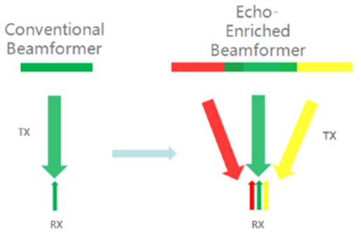
Echo-enriched Beam Forming
Echo-enriched beam former permits the use of traditionally neglected echo signals of adjacent beams to form one more refined and robust imaging beam, providing better out-of-focusimage resolution and deeper image penetration.
Free Xros M™
Gain precise anatomical observation by freely placing sample lines at any angle. Attain better images through a simultaneous display of up to 3 sample lines.
Free Xros CM™
Accurately evaluate myocardial motion at different phases and simultaneously determine myocardial synchronization-- high frame-rate providing you with accurate results.
Auto EF
One intelligent way to analyze 2D echo clips to automatically recognize diastole/systole frames and output EDV/ESV/EF etc. results from Simpson method.
TDI
Tissue Doppler Imaging allows you to quantitatively evaluate local myocardial movement and function, providing complete TDI modes for faster and direct diagnoses.
iScape™
Panoramic imaging gives the user a complete and expanded view of anatomical structures. Couple this with velocity indication and forward/backward scanning abilities, and scanning becomes easier, smoother, and more easily controlled.
ExFOV
Discover better diagnostic information through an extended view of the anatomical structure on all convex and linear probes.
Trapezoid imaging
Discover better diagnostic information through an extended view of the anatomical structure on all linear probes.
Workflow
iZoom™
Gain instant full-screen view with the click of a single key.
iStation™
Mindray’s unique Patient Information Management System allows you to integrate, review, archive, and retrieve patient data effectively.
iTouch™
Gain instant auto image optimization in B, Color, and PW Modes with a single click.
Raw Data
Enables post processing for the stored images for maximum productivity during scanning, including parameter adjustments, adding comments and measurements.
Ergonomics
Innovative Crafted Unit
- Thin Magnesium-alloy body
- 15.6” LED HD monitor with slim design
- Built-in battery providing 90 min scanning time
- High capacity SSD hard drive making patient data safer
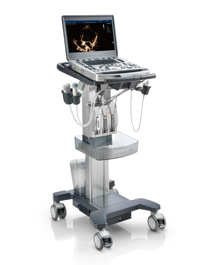
Customized Special Design Trolley
- Inbuilt quick & easy locking system
Green All The Way
- Noiseless system
- Automatic brightness adjustment
- Reliable RoHS certified materials
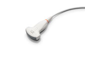
C5-1s
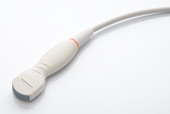
C6-2Gs
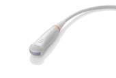
C11-3s
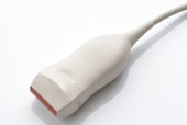
L10-3s
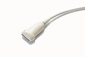
L12-4s
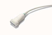
L14-6Ns
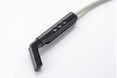
L16-4Hs
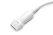
L20-5s
SP5-1s
P10-4s
P7-3s
P8-2s
Related Content