
Display of physiological signal data on the main ultrasound screen
Built-in temperature control module
Secure fixation of transducer with multi-directional and multi-angle adjustment
Anesthesia mask fixation
ECG, respiratory monitoring
- Full rotational and tilt flexibility with adjustable positioning
- Waterproofing, resistance to alcohol and UV disinfection, and anti-oxidation electrodes
- Rectal temperature monitoring
- Temperature management
Precise position adjustments
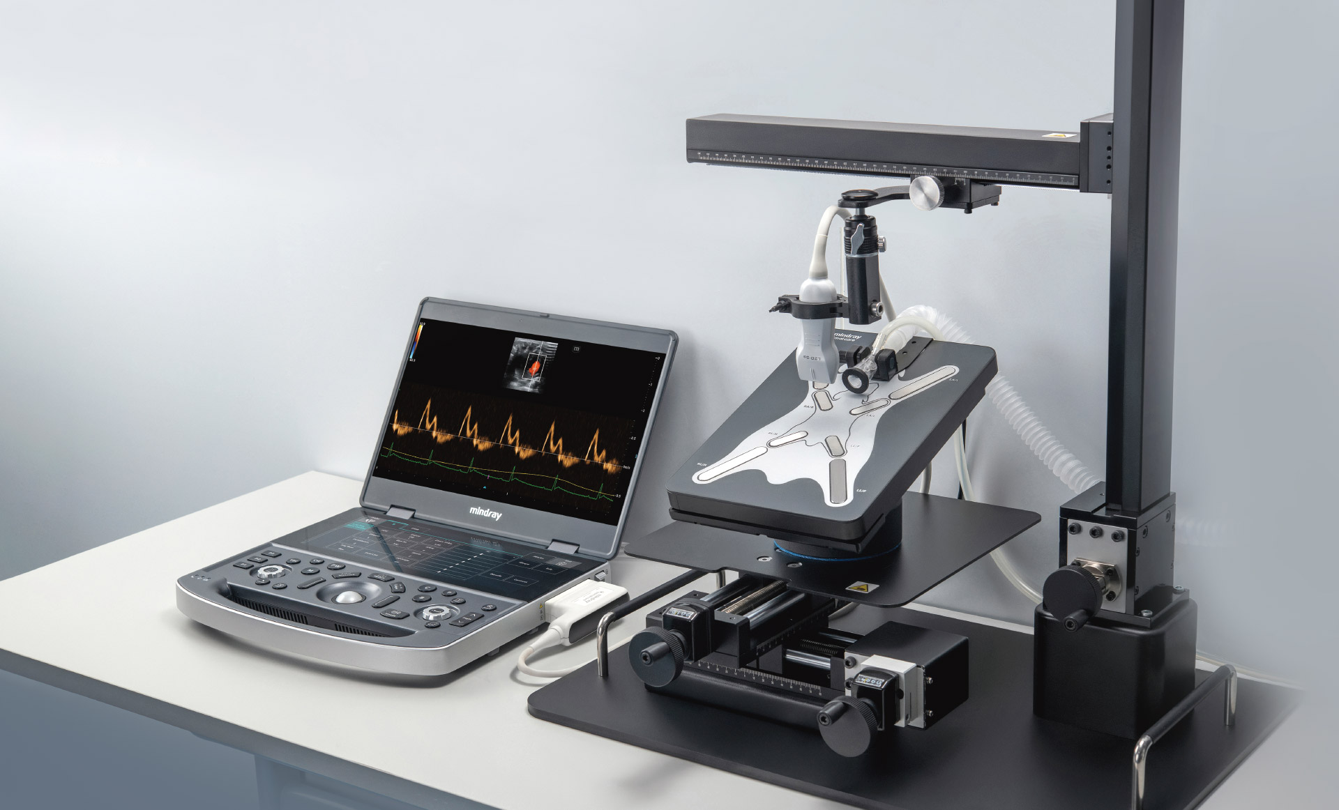
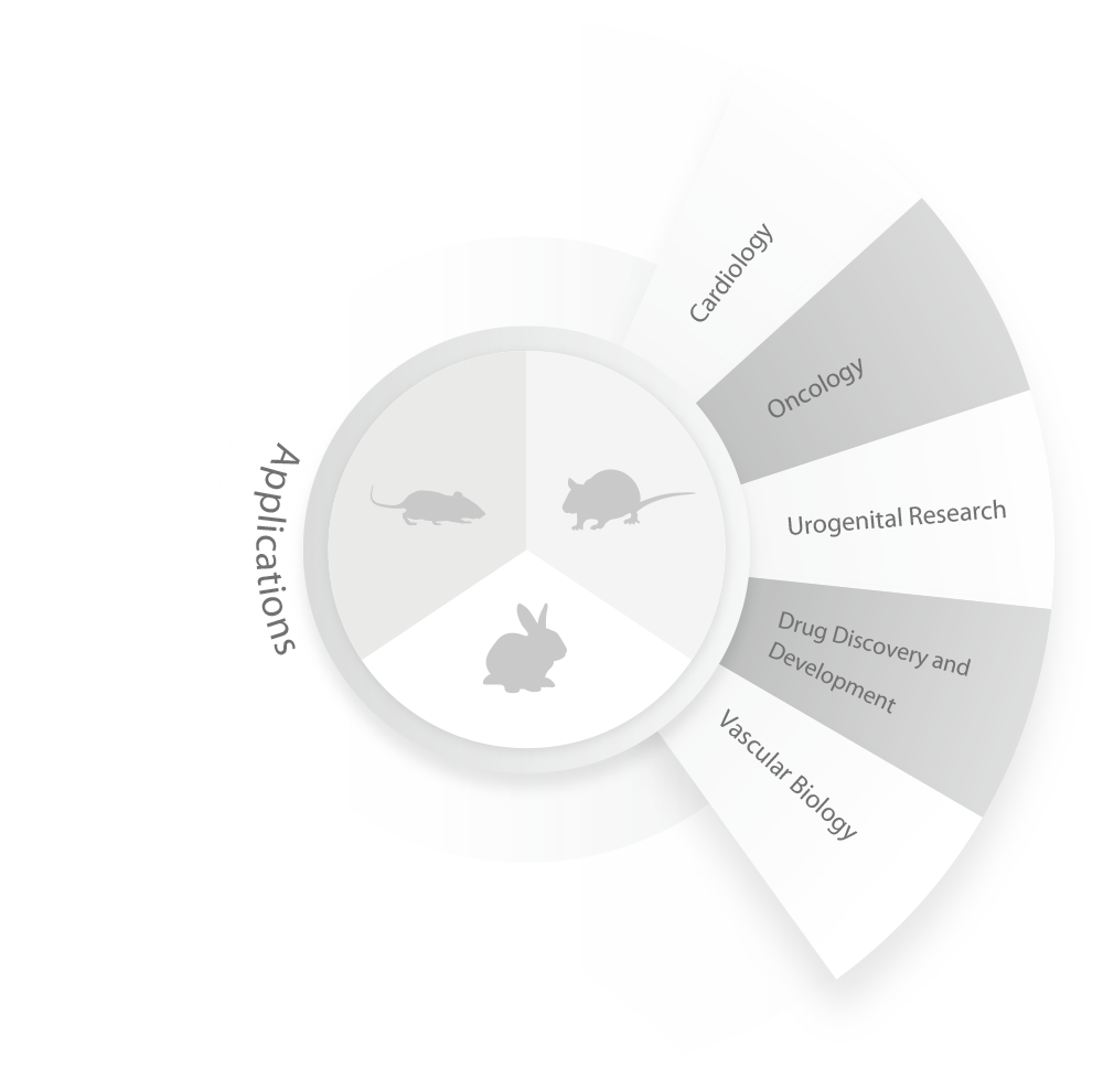
Labus 7 incorporates cutting-edge advancements in ultrasound technology, raising image clarity and measurement precision to new heights. This comprehensive preclinical solution is tailored to meet the demands of cardiovascular, oncology, urology, drug discovery and development research, etc., offering researchers new insights.
Research Excellence Created by Technological Advancements
Research Innovation with Specialized Applications
Research Quality by Precision Engineering
Research Excellence Created by Technological Advancements
ZST+ Imaging Platform
ZONE Sonography® Technology (ZST) is a cutting-edge ultrasound imaging system that employs innovative ultrasound technology. With its advanced hardware and three core technologies—Zone Imaging, Zone Focusing, and Zone Processing—it provides high-resolution imaging suitable for research applications. It supports preclinical studies by offering researchers new perspectives and deeper insights.
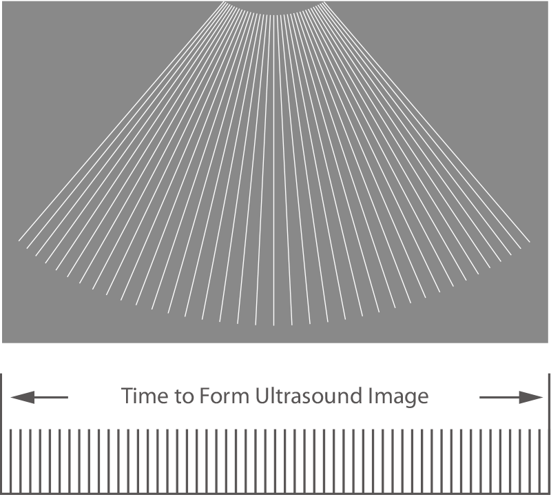
Traditional Beam-forming
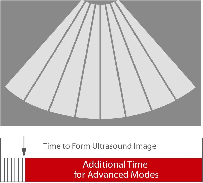
ZONE Sonography™ Technology

Multiple Focusing Based on Traditional Beamforming
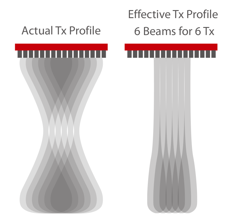
Dynamic Pixel Focusing Based on ZONE Sonography™
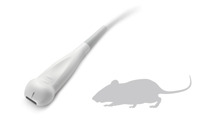
Specialized Research Transducers
Labus 7 features ultra-high-frequency linear array transducers that are compact and lightweight, ideal for use with various laboratory animals such as mice and rats. It delivers exceptional image resolution, capturing fine details with precision. The adjustable imaging parameters ensure highly accurate and reliable experimental results.
L30-8Xs
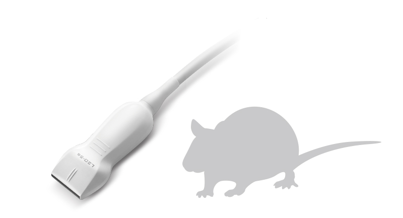
L20-5s
Labus 7 offers a versatile range of transducers for scanning the abdomen and heart in monkeys, dogs, rabbits, and pigs. It also supports advanced imaging modes to meet the requirements for deep tissue imaging in preclinical research.
Research Innovation with Specialized Applications
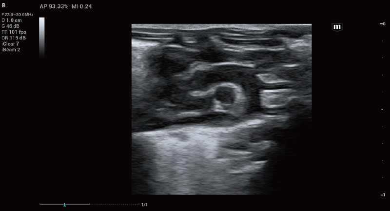
Scanning Modes
Brightness Mode
B-Mode produces two-dimensional grayscale images of tissues, delivering a clear view of their morphology. In research involving laboratory animals, it is used to qualitatively analyze organ structures and sizes, providing essential data for developing and evaluating disease models.
Mouse Aortic Arch
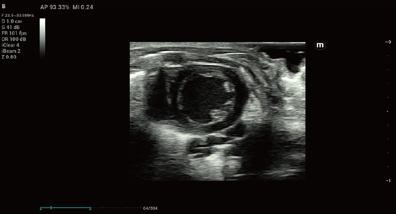
Mouse Left Ventricular Short-axis
Color Doppler Mode
Color Doppler Mode visualizes blood flow and velocity, offering valuable data for hemodynamic studies. It is used to analyze blood flow patterns, vascular network structures, and to investigate microcirculation and overall blood dynamics.
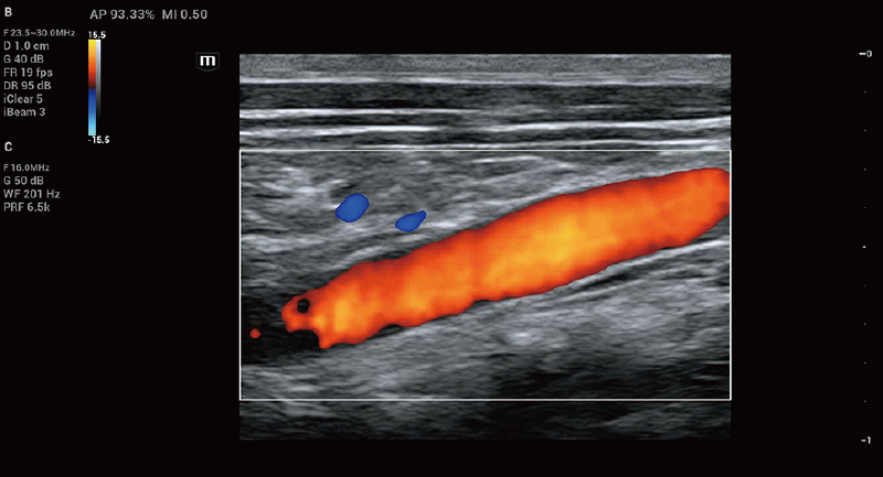
Rat Abdominal Aortic Color Doppler
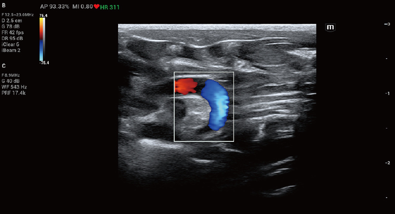
Mouse Aortic Arch Color Doppler
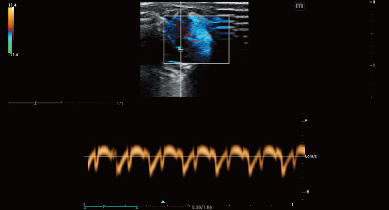
Pulsed Wave Doppler Mode
Pulsed Wave Doppler Mode accurately measures blood flow velocity in specific regions, providing essential data for quantitative hemodynamic studies. It is used in cardiovascular research to measure abnormalities such as stenosis and regurgitation.
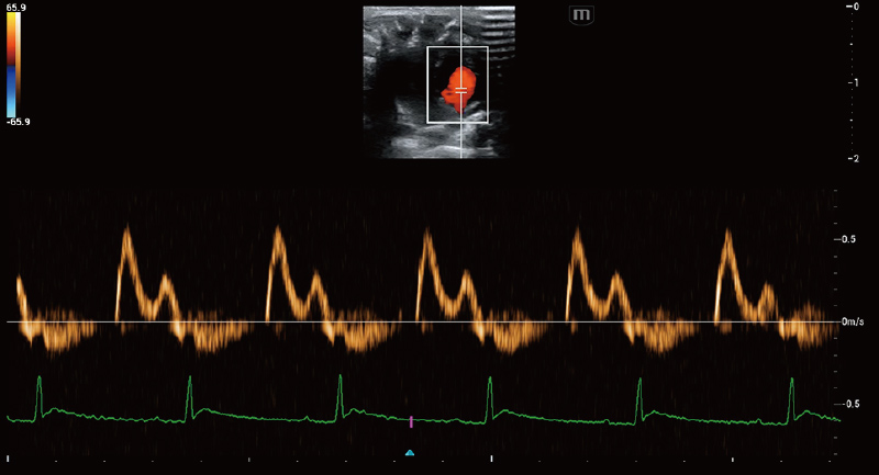
Mouse Mitral Valve PW
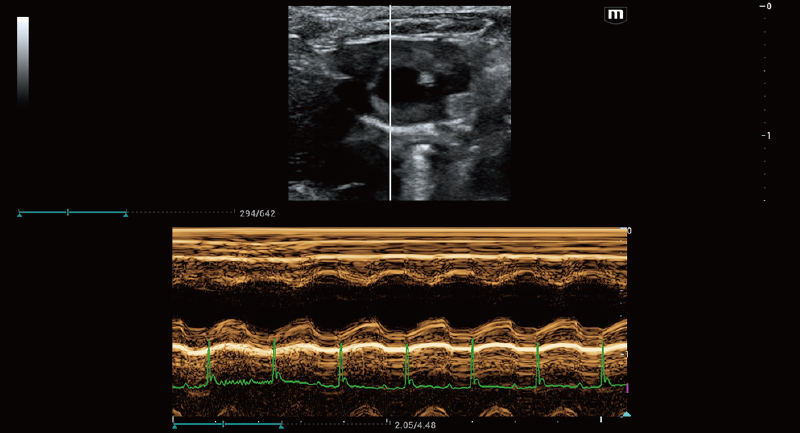
Mouse Left Ventricle M Mode
Motion Mode
M-Mode captures changes in tissue dynamics, offering insights into the motion and function of active organs such as the heart and muscles. It visualizes the movement of heart walls, blood vessel walls, and valve activity.
Preclinical Research Solutions
Cardiovascular Research
Tissue Doppler Imaging Quantitative Analysis
Advanced Tissue Doppler Imaging modes (TVI, TVD, TVM, TEI) allow for synchronized analysis of myocardial motion across up to eight regions of interest (ROIs), offering researchers multidimensional parameters of cardiac tissue movement.
Mouse Mitral Valve Annulus TVD
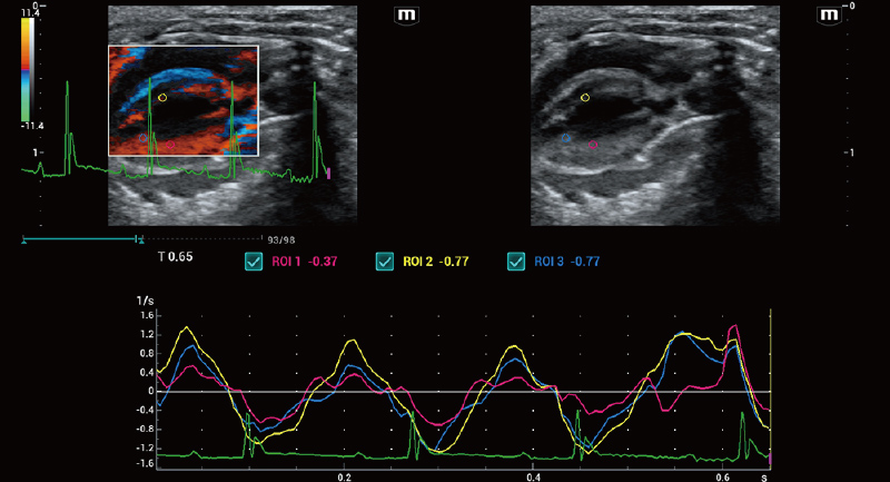
Mouse Left Ventricular TDI QA
Oncology Research
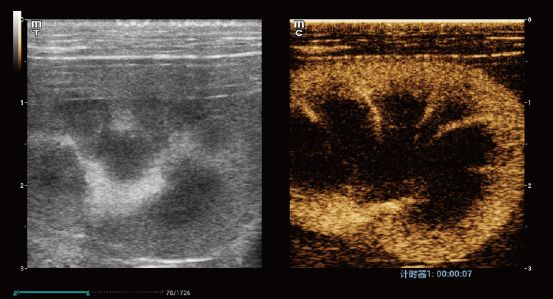
UWN+ Contrast Enhanced Ultrasound
Microbubble contrast agents are used to enhance image contrast, offering clearer views for functional studies of blood vessels and tissues. In studies of tumor angiogenesis and inflammation, it evaluates vascular permeability and blood flow dynamics. UWN+ Imaging System provides quantitative analysis of the entire contrast process, delivering reliable detailed quantitative data.
Rabbit Renal Contrast Imaging
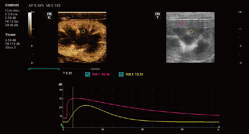
Rabbit Renal Contrast Imaging Quantitative Analysis
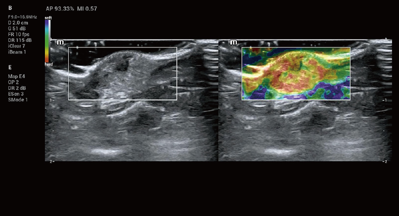
Natural Touch Elastography
It delivers highly sensitive elastography, offering new insights into the physical properties of animal tissues. Its unique Shell analysis feature effectively evaluates the extent of tumor infiltration into surrounding tissues.
Mouse Tumor Elastography

Research Quality by Precision Engineering
Precision engineering of Labus 7 ensures durability and stability in various research settings. With robust data security, it offers researchers a dependable data processing environment, greatly improving the quality and efficiency of their work.

Compact Design

Magnesium-aluminum Alloy Protective Casing

Multi-transducer Expansion

Recycle Bin Accidental Deletion Protection

Data Security Access Control

Built-in High-capacity Hard Drive